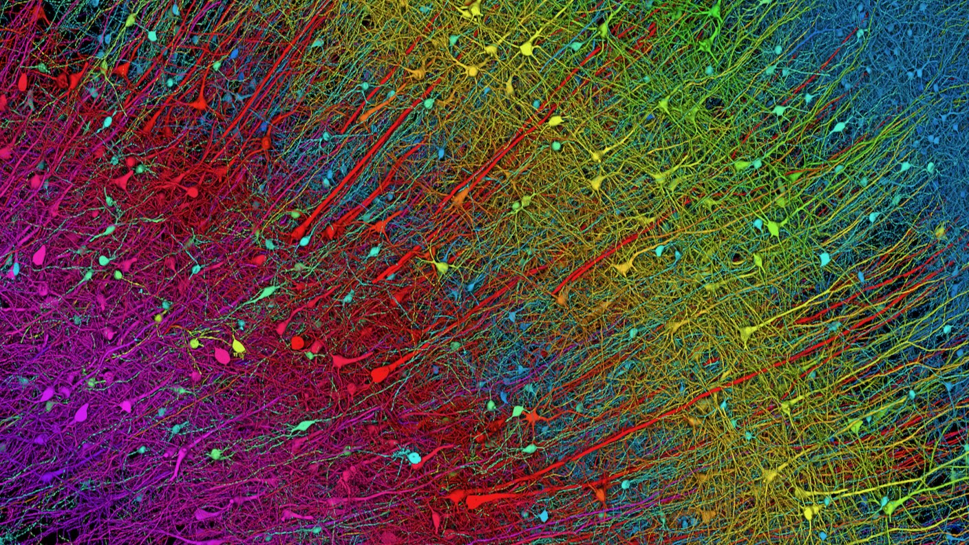
Scientists just published the most detailed map of a cubic millimeter of the human brain. Smaller than a grain of rice, the mapped section of brain includes over 57,000 cells, 230 millimeters of blood vessels, and 150 million synapses.
The project, a collaboration between Harvard and Google, is looking to accelerate connectomics—the study of how neurons are wired together—over a much larger scale.
Our brains are like a jungle.
Neuron branches crisscross regions, forming networks that process perception, memories, and even consciousness. Blood vessels tightly wrap around these branches to provide nutrients and energy. Other brain cell types form intricate connections with neurons, support the brain’s immune function, and fine-tune neural network connections.
In biology, structure determines function. Like tracing wires of a computer, mapping components of the brain and their connections can improve our understanding of how the brain works—and when and why it goes wrong. A brain map that charts the jungle inside our heads could help us tackle some of the most perplexing neurological disorders, such as Alzheimer’s disease, and decipher the origins of emotions, thoughts, and behaviors.
Aided by machine learning tools from Google Research, the Harvard team traced neurons, blood vessels, and other brain cells at nanoscale levels. The images revealed previously unknown quirks in the human brain—including mysterious tangles in neuron wiring and neurons that connect through multiple “contacts” to other cells. Overall, the dataset incorporates a massive 1.4 petabytes of information—roughly the storage amount of a thousand high-end laptops—and is free to explore.
“It’s a little bit humbling,” Dr. Viren Jain, a neuroscientist at Google and study author, told Nature. “How are we ever going to really come to terms with all this complexity?” The database, first released as a preprint paper in 2021, has already garnered much enthusiasm in the scientific field.
“It’s probably the most computer-intensive work in all of neuroscience,” Dr. Michael Hawrylycz, a computational neuroscientist at the Allen Institute for Brain Science, who was not involved in the project, told MIT Technology Review.
Why So Complicated?
Many types of brain maps exist. Some chart gene expression in brain cells; others map different cell types across the brain. But the goal is the same. They aim to help scientists understand how the brain works in health and disease.
The connectome details highways between brain regions that “talk” to each other. These connections, called synapses, number in the hundreds of trillions in human brains—on the scale of the number of stars in the universe.
Decades ago, the first whole-brain wiring map detailed all 302 neurons in the roundworm Caenorhabditis elegans. Because its genetics are largely known, the lowly worm delivered insights, such as how the brain and body communicate to increase healthy longevity. Next, scientists charted the fruit fly connectome and found the underpinnings of spatial navigation.
More recently, the MouseLight Project and MICrONS have been deciphering a small chunk of a mouse’s brain—the outermost area called the cortex. It’s hoped such work can help inform neuro-inspired AI algorithms with lower power requirements and higher efficacy.
But mice are not people. In the new study, scientists mapped a cubic millimeter of human brain tissue from the temporal cortex—a nexus that’s important for memory, emotions, and sensations. Although just one-millionth of a human brain, the effort reconstructed connections in 3D at nanoscale resolution.
Slice It Up
Sourcing is a challenge when mapping the human brain. Brain tissues rapidly deteriorate after trauma or death, which changes their wiring and chemistry. Brain organoids—”mini-brains” grown in test tubes—somewhat resemble the brain’s architecture, but they can’t replicate the real thing.
Here, the team took a tiny bit of brain tissue from a 45-year-old woman with epilepsy during surgery—the last resort for those who suffer severe seizures and don’t respond to medication.
Using a machine like a deli-meat slicer armed with a diamond knife, the Harvard team, led by connectome expert Dr. Jeff Lichtman, meticulously sliced the sample into 5,019 cross sections. Each was roughly 30 nanometers thick—a fraction of the width of a human hair. They imaged the slices with an electron microscope, capturing nanoscale cellular details, including the “factories” inside cells that produce energy, eliminate waste, or transport molecules.
Piecing these 2D images into a 3D reconstruction is a total headache. A decade ago, scientists had to do it by hand. Jain’s team at Google developed an AI to automate the job. The AI was able to track fragments of whole components—say, a part of a neuron (its body or branches)—and stick them back together throughout the images.
In total, the team pieced together thousands of neurons and over a hundred million synaptic connections. Other brain components included blood vessels and myelin—a protective molecular “sheath” covering neurons. Like electrical insulation, when myelin deteriorates, it causes multiple brain disorders.
“I remember this moment, going into the map and looking at one individual synapse from this woman’s brain, and then zooming out into these other millions of pixels,” Jain told Nature. “It felt sort of spiritual.”
A Whole New World
Even a cursory look at the data led to surprising insights into the brain’s intricate neural wiring.
Cortical neurons have a forest-like structure for input and a single “cable” that delivers output signals. Called axons, these are dotted with thousands of synapses connecting to other cells.
Usually, a synapse grabs onto just one spot of a neighboring neuron. But the new map found a rare, strange group that connects with up to 50 points. “We’ve always had a theory that there would be super connections, if you will, amongst certain cells…But it’s something we’ve never had the resolution to prove,” Dr. Tim Mosca, who was not involved in the work, told Popular Science. These could be extra-potent connections that allow neural communications to go into “autopilot mode,” like when riding a bike or navigating familiar neighborhoods.
More strange structures included “axon whorls” that wrapped around themselves like tangled headphones. An axon’s main purpose is to reach out and connect with other neurons—so why do some fold into themselves? Do they serve a purpose, or are they just a hiccup in brain wiring? It’s a mystery. Another strange observation found pairs of neurons that perfectly mirrored each other. What this symmetry does for the brain is also unknown.
The bottom line: Our understanding of the brain’s connections and inner workings is still only scratching the surface. The new database is a breakthrough, but it’s not perfect. The results are from a single person with epilepsy, which can’t represent everyone. Some wiring changes, for example, may be due to the disorder. The team is planning a follow-up to separate epilepsy-related circuits from those that are more universal in people.
Meanwhile, they’ve opened the entire database for anyone to explore. And the team is also working with scientists to manually examine the results and eliminate potential AI-induced errors during reconstruction. So far, hundreds of cells have been “proofread” and validated by humans, but it’s just a fraction of the 50,000 neurons in the database.
The technology can also be used for other species, such as the zebrafish—another animal model often used in neuroscience research—and eventually the entire mouse brain.
Although this study only traced a tiny nugget of the human brain, the atlas is a stunning way to peek inside its seemingly chaotic wiring and make sense of things. “Further studies using this resource may bring valuable insights into the mysteries of the human brain,” wrote the team.
Image Credit: x / x
- SEO Powered Content & PR Distribution. Get Amplified Today.
- PlatoData.Network Vertical Generative Ai. Empower Yourself. Access Here.
- PlatoAiStream. Web3 Intelligence. Knowledge Amplified. Access Here.
- PlatoESG. Carbon, CleanTech, Energy, Environment, Solar, Waste Management. Access Here.
- PlatoHealth. Biotech and Clinical Trials Intelligence. Access Here.
- Source: https://singularityhub.com/2024/05/13/google-and-harvard-map-a-tiny-piece-of-the-human-brain-with-extreme-precision/

