A series of ultrahigh-resolution brain PET images has been selected as the SNMMI Image of the Year. At each of its annual meetings, the Society of Nuclear Medicine and Molecular Imaging chooses an image that represents the most promising advances in the field, with this year’s winner picked from more than 1500 submitted abstracts.
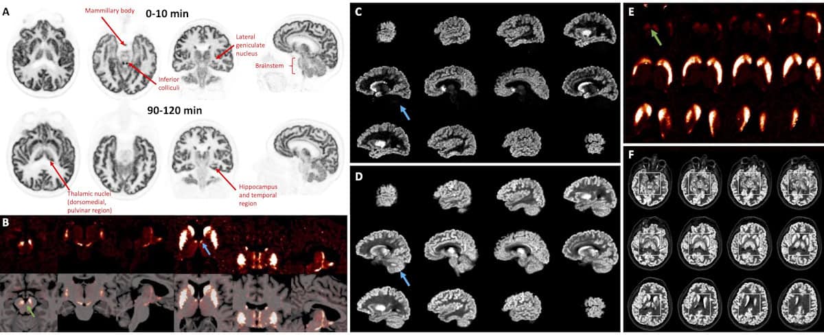
The winning images, recorded by the ultrahigh-performance NeuroEXPLORER human brain PET scanner, highlight targeted tracer uptake in specific brain nuclei (clusters of neurons), providing detailed information on neuronal and functional activity. The technology could dramatically expand the scope of brain PET studies, with potential to advance the treatment of brain diseases.
NeuroEXPLORER was built by a collaboration of researchers from Yale University, the University of California, Davis and United Imaging Healthcare. They designed the scanner to provide ultrahigh sensitivity and ultrahigh spatial resolution, as well as to perform continuous correction for head motion.
“If we are going to get high resolution, we have to deal with patient motion,” Richard Carson from Yale University explained at the recent SNMMI Annual Meeting in Toronto. “But the challenge has always been sensitivity, even with a high-resolution system. In a late frame for a carbon-11 study, you’re really scanning on fumes, it’s hard to generate the quality of images you’d like to really use the resolution that’s possible.”
To achieve the highest possible resolution, ideally better than 2 mm, the team designed a detector micro-block comprising a 4 x 2 array of 1.56 x 3.07 x 20 mm LYSO crystals, read out by four silicon photomultipliers. The 1.5 mm transaxial pitch provides extremely high resolution, while the larger axial pitch is used to read out depth-of-interaction (DOI) data.
The NeuroEXPLORER assembles 20 detector modules, each containing five or six detector blocks (12 × 12 microblocks), in a cylindrical ring with a diameter of 52.4 cm. The scanner’s high sensitivity is enabled by having a long (49.5 cm) axial field-of-view, building on pioneering work from UC Davis on the EXPLORER system. Motion correction is performed using a Vicra tracking system with a built-in camera that collects data from the face for markerless motion tracking.
In performance tests, the NeuroEXPLORER demonstrated a sensitivity of 46 kcps/MBq at the centre, a transverse spatial resolution of less than 1.4 mm with OSEM (ordered-subset expectation-maximization) reconstruction, and a DOI resolution of less than 4 mm. The scanner also performs time-of-flight measurements with an average resolution of 236 ps. The team provide detailed system characteristics in the Journal of Nuclear Medicine.
Exceptional images
In their latest study, presented by Carson at the SNMMI Annual Meeting, the researchers compared human brain images from the NeuroEXPLORER with images from the current state-of-the-art scanner, the High Resolution Research Tomograph (HRRT).
The team scanned healthy volunteers, using both PET systems on different days, after administration of five different radiotracers. The tracers target various features in the brain, including synaptic density (imaged with 18F-SynVesT-1), dopamine receptors (11C-PHNO) and transporters (18F-FE-PE2I), and glucose metabolism (18F-FDG). Carson also shared some late-breaking data using the tracer 18F-Flutabine.
Overall, the team observed a dramatic improvement in the resolution and quality of NeuroEXPLORER images compared with those recorded by the HRRT. Carson presented a series of examples to the SNMMI audience.
NeuroEXPLORER images using 18F FDG, for instance, visualized extremely small substructures with much higher contrast than corresponding HRRT scans. Time activity curves in the caudate and thalamus quantified that NeuroEXPLORER showed higher standard uptake values (SUV) than HRRT. Likewise, late images of synaptic density, recorded 60–90 min postinjection, showed higher contrast and greater SUV with the NeuroEXPLORER than the HRRT scanner.
Carson also presented binding potential images using the dopamine receptor tracer 11C-PHNO. While noise is higher with a 11C-based tracer, NeuroEXPLORER scans showed higher values than the HRRT images. “This was one of the really good days,” he said. “We saw two hotspots in the anterior thalamus, we’re not sure exactly which subnucleus within there, but we’re seeing beautiful hotspots consistent with high dopaminergic intervention in those regions. If you look at the HRRT images, it’s there, but not something you could commit to.”
Carson noted that the NeuroEXPLORER’s long axial field-of-view enables the scanner to also image the carotid arteries in the neck. In addition, the team is using 18F-FDG to image head-and-neck tumours, for example to visualize lymph nodes in the neck. “This is an exciting opportunity in terms of being able to see much smaller tumours,” he said.
The team’s ongoing projects include optimization of the reconstruction parameters, further improving the camera for motion correction, and fully characterizing the resolution of the PET images using brain phantoms with variable contrast.
Deep transfer learning detects six different cancers on PET/CT scans
As well as imaging of small brain structures with targeted pharmaceuticals, future studies will also include more imaging in the spinal cord, and looking at the dynamics of neurotransmitter release. “One exciting part to think about doing is, because of the sensitivity, we can drop the radiation dose and begin to scan adolescents, for example with autism or schizophrenia,” said Carson.
“The dramatic improvement in resolution and overall quality of the NeuroEXPLORER images compared to the HRRT images is clear,” says Heather Jacene, chair of SNMMI’s Scientific Program Committee, in a press statement. “The NeuroEXPLORER has the potential to be a gamechanger in research for conditions such as Alzheimer’s disease, Parkinson’s disease, epilepsy and mental illnesses.”
- SEO Powered Content & PR Distribution. Get Amplified Today.
- PlatoData.Network Vertical Generative Ai. Empower Yourself. Access Here.
- PlatoAiStream. Web3 Intelligence. Knowledge Amplified. Access Here.
- PlatoESG. Carbon, CleanTech, Energy, Environment, Solar, Waste Management. Access Here.
- PlatoHealth. Biotech and Clinical Trials Intelligence. Access Here.
- Source: https://physicsworld.com/a/snmmi-image-of-the-year-visualizes-the-brain-as-never-before/

