
The Annual Meeting of the Society of Nuclear Medicine and Molecular Imaging (SNMMI), held this week in Chicago, saw researchers showcase the latest technology developments, clinical advances and new radiopharmaceuticals for imaging and treatment of disease. Among the device innovations highlighted at the meeting, investigators presented some novel instrumentation designed to improve the performance of PET.
‘Outsert’ device boosts PET performance
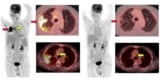
Researchers at Washington University in St. Louis are using a new technology called “augmented whole-body scanning via magnifying PET” (AWSM-PET) to enhance the resolution and sensitivity of clinical whole-body PET/CT imaging. The cost-effective technology uses high-resolution add-on detectors that simultaneously scan a patient during a standard whole-body PET scan.
“Whole-body PET/CT imaging is broadly used for cancer staging and restaging and to evaluate patients’ response to treatment interventions; however, its diagnostic accuracy is compromised when the lesions are very small or exhibit weak signals,” explained Yuan-Chuan Tai, who presented the study at the meeting. “Our novel AWSM-PET prototype helps to tackle two of the key limitations in whole-body PET imaging: image resolution and overall system sensitivity.”
The AWSM-PET technology utilizes two high-resolution PET detectors that are placed outside of a scanner’s axial imaging field-of-view, which the researchers call an “outsert” device. Each outsert panel comprises 32 LSO crystal arrays, each containing 30×30 elements (0.97×0.97×10.0 mm each). The device simultaneously acquires high-resolution PET data while a patient undergoes whole-body PET, requiring no additional scanning time. The team also developed custom reconstruction and correction algorithms to jointly reconstruct the data.
To test their technology, the researchers used a prototype AWSM-PET device implemented on a Siemens Biograph Vision PET/CT scanner. They imaged cylindrical phantoms containing tumour inserts of varying size, observing a clear improvement in image resolution when data from the outsert device were included. They note that the outsert detectors exhibited excellent spatial, energy and timing resolution, with a system-level coincidence resolving time of 217 ps – suitable for time-of-flight applications.
“The additional high-resolution data from the AWSM-PET device can enhance the overall image resolution and reduce statistical noise,” noted Tai. “The potential improvement in diagnostic accuracy of clinical whole-body PET/CT may benefit cancer patients.”
Tai and colleagues plan to start a pilot human imaging trial later this year at the Washington University School of Medicine in St. Louis. The study will compare the diagnostic accuracy of AWSM-PET versus standard-of-care whole-body PET/CT.
A quantum leap in brain PET resolution
High spatial resolution is essential for effective brain PET, to enable visualization and characterization of biological processes occurring in small cerebral structures. With this aim, a research team headed up at the Université de Sherbrooke in Canada has developed an ultrahigh-resolution (UHR) brain PET scanner. The system enabled characterization of previously indistinguishable brain regions that are involved in conditions such as Alzheimer’s disease, depressive and visual attention disorders, and tinnitus.
“Up until now, PET has been useful for the study of neurological phenomena and for diagnostic purposes, but its potential has been somewhat limited by the poor spatial resolution of current PET systems,” explained master’s student Vincent Doyon, who shared the first brain images acquired with the new scanner.
The UHR scanner features pixelated detectors with one-to-one coupling between the scintillators and photodetectors. This results in a spatial resolution of 1.12 mm – more than twice as good as a the current state-of-the-art for brain PET imaging. Having demonstrated the UHR scanner’s imaging capabilities using resolution phantoms and preclinical studies, the researchers have now investigated its potential for human brain imaging.
Four patients underwent a clinical 18F-FDG PET exam on a whole-body PET scanner for 10 to 20 min, followed by a 30- to 60-min brain scan on the UHR scanner. The team reconstructed the UHR images using an OSEM algorithm, with CT-based attenuation and scatter correction, and then performed region identification and calculated standardized uptake values relative to the cerebellum.

The UHR images clearly identified several regions of the brain (particularly in the brainstem) that could not be resolved by the whole-body scanner, including many structures that had never been seen before using FDG-PET. The team also observed hypermetabolic regions along the cortical surface in the UHR images that were hardly perceived with the whole-body PET scanner.
SNMMI Annual Meeting highlights advances in nuclear medicine
Doyon pointed out that while standard PET images usually visualize the thalamus as a uniform mass, the UHR images could be segmented into smaller thalamic nuclei. This is a promising finding, he explained, as these nuclei are involved in many physiological functions and affected by diseases in specific ways.
“The UHR scanner is a quantum leap for PET image resolution,” said Doyon. “Proper visualization of brainstem nuclei will provide the ability to detect early changes associated with many diseases and offer a potential avenue for early diagnosis. This will impact both research and clinical settings.” The first UHR prototype is now fully operational and being used for research at the Sherbrooke Molecular Imaging Center.
- SEO Powered Content & PR Distribution. Get Amplified Today.
- PlatoData.Network Vertical Generative Ai. Empower Yourself. Access Here.
- PlatoAiStream. Web3 Intelligence. Knowledge Amplified. Access Here.
- PlatoESG. Automotive / EVs, Carbon, CleanTech, Energy, Environment, Solar, Waste Management. Access Here.
- BlockOffsets. Modernizing Environmental Offset Ownership. Access Here.
- Source: https://physicsworld.com/a/innovative-devices-ramp-the-resolution-of-pet-imaging/

