
Dynamic positron emission tomography (PET) is a medical imaging technique that tracks both the spatial and temporal pattern of radiotracer uptake. The approach provides information that can’t be gained by conventional static PET, such as perfusion and diffusion information and pharmacokinetic parameters that help scientists understand how long a tracer stays somewhere in the body, where the tracer came from, and where it goes afterward.
Dynamic PET has been used in cardiology, treatment response assessments, theranostics applications, drug discovery research, diagnosis and research on neurodegenerative diseases, and more. It can generate multiple types of PET images with complementary information in a single imaging session, such as influx rate, distribution volume and conventional standardized uptake value (SUV)-equivalent images.
Yet, dynamic PET to date is largely confined to academic research centres.
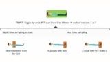
That’s in part because to create a dynamic PET image series, scientists need to obtain an image-derived arterial input function – the radiotracer concentration in the patient’s arterial blood plasma as a function of time. The current gold standard for arterial blood sampling is an invasive and expensive procedure that requires many clinical resources, including anaesthesiologists and surgical space.
Several research groups, including Shirin Abbasinejad Enger’s team at the Jewish General Hospital, McGill University, are working to change that.
“We’re trying to simplify this clinical workflow by removing the invasive arterial blood sampling step,” says Liam Carroll, a PhD student in Enger’s group. “We can measure this radiation by placing a radiation detector on a patient’s wrist, which gives you the same information you would normally collect with arterial blood sampling.”
Carroll and Enger have developed a cost-effective radiation detector that acquires the arterial input function non-invasively. Patients who have been injected with a radiotracer place their wrist on the detector, which measures the radiation escaping from the patient’s wrist. Then, an algorithm is used to derive the arterial input function from the detector data.
The researchers introduced a detector prototype in 2020. Since then, they’ve been working to optimize detector size and geometry and develop a clinical data analysis chain to accurately separate arterial and venous components of arterial input functions. A recent study reported in Medical Physics presents their latest results.
“We would like to provide any centre that already has a PET scanner with an affordable detector that they can use for dynamic PET scanning, if needed,” says Enger. “So one of the first decisions we made was that this detector needs to be affordable such that clinics with less resources can also acquire dynamic PET images, because any PET scanner can be used for dynamic imaging.”
Detectors designed by other research groups, Enger says, are larger and less cost-effective. Several approaches use block crystal scintillators, while others arrange detector elements in rings, similar to a miniature PET detector. While these detectors may have higher detection efficiencies, they also may cause artefacts when placed in the field-of-view of the PET scanner, she explains. Placing detectors elsewhere on the body, such as the leg, still may not be suitable for whole-body dynamic PET scanning.
Carroll and Enger’s detector couples silicon photomultipliers to low-density scintillating fibres surrounded by a 3D-printed plastic shell. The detector has two layers of narrow scintillating fibres, which maximizes the total and intrinsic efficiency of the detector while keeping manufacturing costs and electronic complexity relatively low. Detector efficiency in the first row of scintillators ranges from approximately 53% to 84% for positrons and 51% to 52% for annihilation photons (total efficiency ranges from approximately 62% to 74%) across four radioisotopes used in a variety of dynamic PET applications (fluorine-18, carbon-11, oxygen-15 and gallium-68).
Dynamic PET edges closer to clinical prime time
The clinical data analysis pipeline uses an expectation maximization maximum likelihood algorithm. It includes counts from 511 keV annihilation photons, Carroll says, because positrons emitted from low-energy positron emitters do not reach the detector. The research team is planning to incorporate radial artery and wrist diameters obtained during ultrasound imaging to improve their data analysis pipeline.
Carroll and Enger have already built and tested a prototype detector and have founded a start-up, BetaScint Imaging, to commercialize it. They are currently preparing to validate the detector’s performance in vivo in patient clinical trials, which will begin in early next year.
“Dynamic PET studies right now are benefiting a very small percentage of patients who need it,” Enger says. “Our hope is that more patients will benefit from this imaging technique in the future.”
- SEO Powered Content & PR Distribution. Get Amplified Today.
- Platoblockchain. Web3 Metaverse Intelligence. Knowledge Amplified. Access Here.
- Source: https://physicsworld.com/a/compact-radiation-detector-could-expedite-the-use-of-dynamic-pet/

