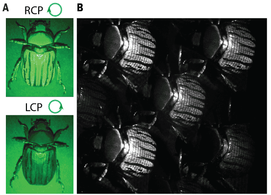
The polarization of light scattered off an object provides a treasure trove of information. Techniques that image this polarization are often overlooked, however, because they are difficult to implement outside a laboratory setting. Researchers at Harvard University, US, have addressed this problem by developing a compact, single-shot polarization imaging system based on a technique called Mueller matrix imaging. The new system replaces many elements in traditional systems with nano-engineered metasurfaces, and its developers say it might find applications in biomedical imaging as well as fundamental research.
The colour of an object – that is, the frequency of light that scatters off it – generally depends on the colour of light incident on it. In the same way, the polarization of the light scattered off an object depends on the polarization of light that illuminates it. Mueller matrix imaging works by controlling the polarization of this incident light, and it is currently the most complete method for imaging polarization, revealing information that would be impossible to obtain using traditional techniques. It is difficult to implement in practice, however, because it requires complex apparatus that uses multiple rotating plates and polarizers to capture a sequence of images that are then combined to produce a matrix.
A much simpler system
The new system, developed by Federico Capasso and colleagues in the electrical engineering department at Harvard’s John A Paulson School of Engineering and Applied Sciences (SEAS), is much simpler. At its heart are two metasurfaces – artificially engineered ultrathin films made from arrays of tiny dielectric structures. These structures, which behave somewhat like atoms, are termed “meta-atoms” and are separated from each other by distances smaller than the wavelengths of the incoming light.
As light passes through the two metasurfaces, its properties – such as its amplitude, phase and polarization – change. “The first surface generates what’s known as polarized structured light, in which the polarization is designed to vary spatially in a unique pattern,” explains Aun Zaidi, who was involved in the work as a PhD student in Capasso’s laboratory and is now an optical scientist and engineer at Apple. “When this polarized light reflects off or transmits through the object being illuminated, the polarization of the profile beam changes. This change is captured and analyzed by the second metasurface to construct the final image – in a single shot.”
The nanoengineered metasurfaces greatly simplify the system’s design, Zaidi adds. Indeed, the system is free of any moving parts or bulk polarization optics and will allow for applications in real-time medical imaging, materials characterization, machine vision and target detection, to name but a few. “Our new compact system might even unlock the vast potential of polarization imaging for a range of existing and new applications, including augmented and virtual reality systems, for example, for facial recognition in smartphones and eye-tracking,” he says.
Other potential applications
In the near term, the new technique, which the team describe in Nature Photonics, could prove useful in applications that require both compact and single-shot polarization imaging. “In biomedicine, these include imaging live tissue samples in microscopy, polarimetric endoscopic imaging, retinal scanning and non- or minimally-invasive imaging of cancerous tumours,” Zaidi tells Physics World.

Metasurfaces simplify optical sensing systems
Thanks to its superior time resolution and flexibility, the technique might also be employed to generate large datasets for training neural networks in machine learning classification applications, he adds. “Beyond these, it might also be important for fundamental science, such as in the detection of the time-varying birefringence of the vacuum in the presence of intense electric and magnetic fields (as theorized by quantum electrodynamics) for the study of three-dimensional polarization states of light, and in research on both short-wavelength (X-ray) and long-wavelength (terahertz) polarimetry,” Zaidi says. “We plan to expand our work into these exciting new directions.”
- SEO Powered Content & PR Distribution. Get Amplified Today.
- PlatoData.Network Vertical Generative Ai. Empower Yourself. Access Here.
- PlatoAiStream. Web3 Intelligence. Knowledge Amplified. Access Here.
- PlatoESG. Carbon, CleanTech, Energy, Environment, Solar, Waste Management. Access Here.
- PlatoHealth. Biotech and Clinical Trials Intelligence. Access Here.
- Source: https://physicsworld.com/a/metasurfaces-make-a-single-shot-polarization-imaging-system/


