Monitoring changes in cerebral blood flow provides a valuable tool for diagnosing brain disorders. Transcranial Doppler (TCD) ultrasound offers a low-cost, non-invasive way to evaluate blood flow in the brain, but the rigid design and manual operation of conventional systems make continuous use impractical. And because TCD probes generally employ a single transducer or linear transducer array, they can only image part of the complex 3D network of cerebral arteries.
To address these limitations, Sheng Xu and colleagues at the University of California San Diego have developed a wearable ultrasound patch for hands-free volumetric imaging of cerebral arteries and long-term monitoring of blood flow in the brain, describing the device in Nature.
“The continuous monitoring capability of the patch addresses a critical gap in current clinical practices,” says co-first author Sai Zhou in a press statement. “Typically, cerebral blood flow is monitored at specific times each day, and those measurements do not necessarily reflect what may happen during the rest of the day. There can be undetected fluctuations between measurements. If a patient is about to experience an onset of stroke in the middle of the night, this device could offer information that is crucial for timely intervention.”
Device design and functionality
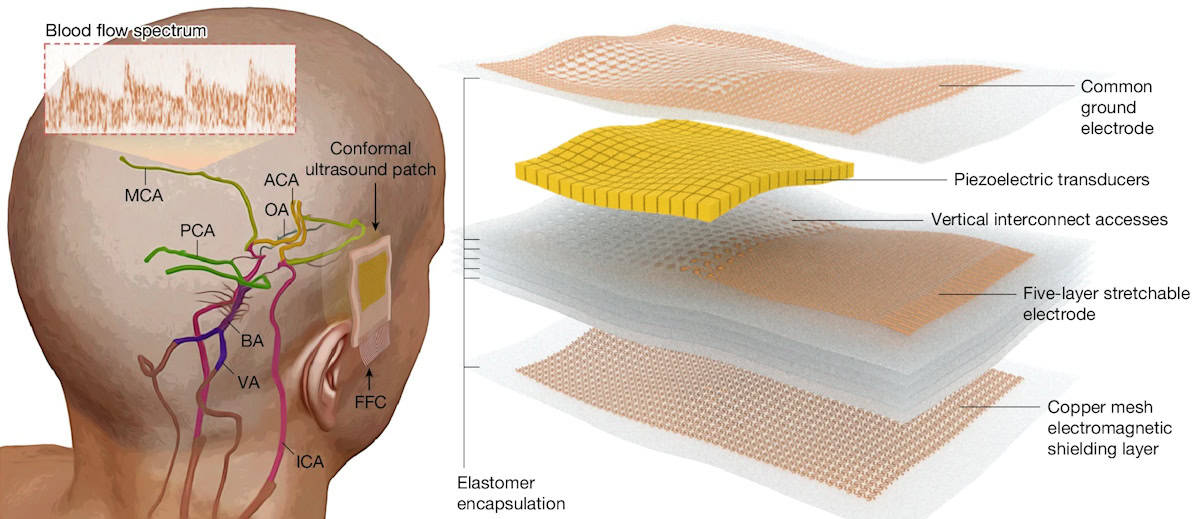
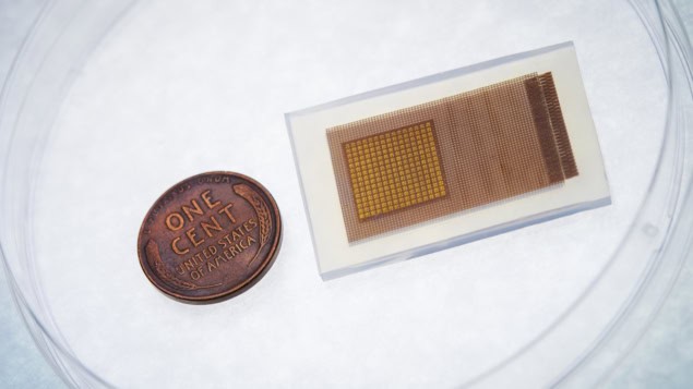
The postage stamp-sized ultrasound patch comprises a 16 x 16 array of piezoelectric transducers with a 2-MHz centre frequency, chosen to minimize attenuation and phase aberration as the signal passes through the skull. The researchers incorporated a layer of copper mesh to provide electromagnetic shielding and increase the signal-to-noise ratio, and encapsulated the entire device in a waterproof silicone elastomer. During use, the patch is linked via shielded cables to a commercial ultrasound machine.
The cerebral vasculature is a complex network comprising multiple arteries, most of which are deep and widely distributed inside the brain. To examine this entire network in 3D, the patch performs ultrafast ultrasound imaging (3000 images per second) and simultaneously emits five diverging waves at different angles, extending the imaging field to about 60 x 60 mm at 50 mm depth.
When attached to a volunteer’s temporal window (in front of the ear), the patch could insonate the terminal internal carotid artery (TICA), which delivers blood from the neck to the major arteries in the brain, as well as the anterior cerebral arteries (ACA), middle cerebral arteries (MCA) and posterior cerebral arteries (PCA), which deliver blood to most of the four brain lobes.
Following signal acquisition, the researchers used customized algorithms to process the data and reconstruct a volumetric power Doppler image of this large vascular network. This 3D reconstruction can then be used to focus the ultrasound beam on targeted arterial sections for long-term monitoring of blood flow spectra.
To validate the performance of the ultrasound patch, the researchers compared blood flow measurements with those recorded by a conventional TCD probe. They recorded blood flow velocities of 10 arterial segments in 36 healthy volunteers, using four transcranial windows (temporal, orbital, submandibular and suboccipital) to target different arterial segments.
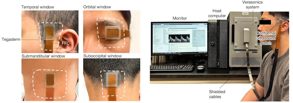
The mean differences between blood flow velocities measured by the ultrasound patch and the conventional TCD probe were −1.51±4.34, −0.84±3.06 and −0.50±2.55 cm/s for peak systolic, mean flow and end diastolic velocity, respectively – demonstrating good agreement between the two devices.
Tracking blood flow changes
Next, the team used the ultrasound patch while participants conducted various activities that modulate blood flow in specific cerebral arteries.
When performing a handgrip, for example, contraction of the forearm muscles activates the sympathetic nervous system, increasing blood supply to the brain and blood flow velocity in the contralateral MCA. As the volunteer began a handgrip, the left MCA mean flow velocity increased rapidly then slowly plateaued at around 119% baseline velocity when the handgrip was maintained. Immediately after releasing the handgrip, the velocity swiftly decreased.
The researchers carried out similar measurements while volunteers performed the Valsalva manoeuvre, word generation and visual stimulation. In all cases, the patch recorded cerebral blood flow changes in accordance with the activities being performed.
Such transient changes in cerebral blood flow can be impaired or inhibited by brain disease or disorders, indicating the potential clinical benefit the ultrasound patch offers for diagnostic applications.

Wearable ultrasound sensor provides continuous cardiac imaging
Finally, the researchers demonstrate the potential for long-term surveillance using the ultrasound patch, monitoring cerebral blood flow spectra in the MCA in a participant continuously for 4 h. The patch identified a cascade of B waves (slow oscillations in cerebral blood flow velocity that are related to waste removal in the brain and disease recovery) when the participant felt drowsy.
“Next, we will collaborate with clinicians to recruit pregnant participants with different conditions, and validate the performance and efficacy of our device compared with handheld clinical ultrasound devices,” Xu tells Physics World. “Additionally, we will try to integrate customized electronics to make the patch wireless, so that people can wear it without any cable tethering.”
- SEO Powered Content & PR Distribution. Get Amplified Today.
- PlatoData.Network Vertical Generative Ai. Empower Yourself. Access Here.
- PlatoAiStream. Web3 Intelligence. Knowledge Amplified. Access Here.
- PlatoESG. Carbon, CleanTech, Energy, Environment, Solar, Waste Management. Access Here.
- PlatoHealth. Biotech and Clinical Trials Intelligence. Access Here.
- Source: https://physicsworld.com/a/ultrasound-patch-continuously-tracks-blood-flow-in-the-brain/

