
Photon-counting CT (PCCT), an advanced medical imaging technique that measures the energy of each individual X-ray photon, is known to improve cardiovascular CT imaging in adults. Now, a study from Germany published in Radiology shows that PCCT similarly improves the image quality for newborn babies and infants suspected of having congenital heart defects.
Congenital heart defects, the most common type of birth defect, are usually diagnosed using pre- and post-natal ultrasound imaging. But ultrasound does not provide sufficient image quality to make a comprehensive assessment of individual anatomy, especially in complex malformations in infants. If surgery is required, CT and MRI can be employed for treatment planning; but both have limitations when used with babies.
Researchers at the RWTH Aachen University Hospital hypothesized that first-generation PCCT might produce better quality images than third-generation energy-integrating dual-source CT (DSCT) scans. PCCT offers the advantages of converting X-ray photons directly into electrical current, which may avoid signal loss at the detector. This should reduce electronic noise, thus increasing the signal-to-noise ratio (SNR) and contrast-to-noise ratio (CNR) and/or enable imaging with reduced radiation dose.
“Infants and neonates with suspected congenital heart defects are a technically challenging group of patients for any imaging method, including CT,” comments principal investigator Timm Dirrichs. “There is a substantial clinical need to improve cardiac CT of this vulnerable group. It’s essential to carefully map the individual cardiac anatomy and possible routes of surgical intervention using the highest possible diagnostic standards.”
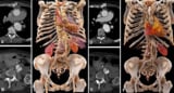
Dirrichs and colleagues conducted a prospective study comparing image quality and radiation exposure of 83 infants with suspected congenital heart defects who underwent contrast-enhanced DSCT (using Siemens Healthineers’ Somatom Force), 30 who underwent contrast-enhanced PCCT (using the Naeotom Alpha) and one infant who had both scans.
For each image, the researchers calculated the SNR and CNR in standardized regions-of-interest placed in the descending aorta and subcutaneous fat tissue. They also estimated the effective radiation exposure using CT dose index and dose–length product. Two radiologists, one paediatric cardiologist and one paediatric cardiac surgeon independently rated the images on a five-point scale for sharpness, overall visual contrast, delineation of vessels, motion artefacts, ring artefacts, quality of 3D reconstructions and overall image quality.
In all but one of the PCCT scans (97%), the CT images were deemed of diagnostic quality, compared with 77% of the DSCT scans. The sole non-diagnostic PCCT exam was the result of a missed contrast agent bolus. The 19 non-diagnostic DSCT exams had prohibitively low SNR and CNR, image artefacts or inadequate contrast agent timing.
Quantitative assessment showed that both SNR and CNR were significantly higher for PCCT images, with a mean SNR of 46.3 and a CNR of 62.0, compared with 29.9 and 37.2, respectively, for DSCT. The mean effective radiation doses were similar: 0.50 mSv for PCCT and 0.52 mSv for DSCT.
Finally, in terms of overall image quality, PCCT significantly outperformed DSCT. The radiology team rated 40% of PCCT images as excellent and 47% as good, compared with 4% and 32%, respectively, for the DSCT images. The team reports that PCCT also outperformed DSCT in of all the other comparative categories.
Photon-counting detectors lower contrast-related risks in CT angiography
The researchers point out that the results of their PCCT assessment are conservative, because the PCCT cohort had a younger median age, size and weight than the DSCT cohort. They attribute this to the fact that after a PCCT scanner became available, paediatric cardiac surgeons referred increasingly younger patients to them due to the image quality being obtained.
The investigators conclude that photon-counting CT offers better cardiovascular imaging quality than dual-source CT at a similar radiation dose in children with suspected heart defects. They believe that PCCT could also be useful for detailed tissue characterization, iodine mapping and creation of 3D models. “High SNR and CNR of the underlying cross-sectional CT images are crucial to delineate small cardiac structures on 3D models or virtual reality models,” they write. “The resulting holograms or 3D prints are increasingly required by paediatric cardiology surgeons for every surgery.”
- SEO Powered Content & PR Distribution. Get Amplified Today.
- PlatoData.Network Vertical Generative Ai. Empower Yourself. Access Here.
- PlatoAiStream. Web3 Intelligence. Knowledge Amplified. Access Here.
- PlatoESG. Automotive / EVs, Carbon, CleanTech, Energy, Environment, Solar, Waste Management. Access Here.
- BlockOffsets. Modernizing Environmental Offset Ownership. Access Here.
- Source: https://physicsworld.com/a/photon-counting-ct-improves-cardiac-imaging-in-infants-with-heart-defects/

